
Bone histology - Notes & Video

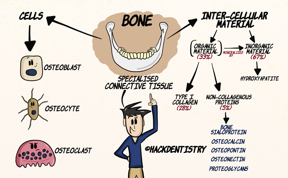
This video covers both the basics of bone histology and the structure of the alveolar bone. However, since this topic can be overwhelming I have split the whole video topic into two notes - one covering bone histology and the other covering the structure of the alveolar bone. This note covers the histology of bone.Bone is a specialised form of connective tissue that consists of cells and intercellular material.
Osteoblasts, osteocytes and osteoclasts make up the cells of the bone, while the inter-cellular material is made of organic and inorganic material.
By dry weight, bone comprises of 33% organic matrix, with 28% representing type I collagen, and non-collagenous proteins like bone sialoprotein, osteocalcin, osteopontin, osteonectin and proteoglycans making up 5%.
The organic matrix is mineralized by hydroxyapatite (inorganic material), which comprises the rest of the 67% of bone by dry weight.
Though bone can be classified into different types based on its shape as long (eg.tibia, humerus), short (eg.carpals, tarsals), flat (eg.sternum,scapula) or irregular(vertebrae, maxilla); most bones are essentially made of two tissue patterns called
a) compact or cortical bone and a
b) fine network of cancellous bone or spongy/trabecular bone.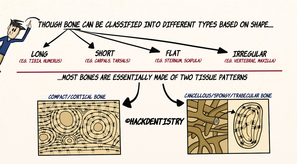
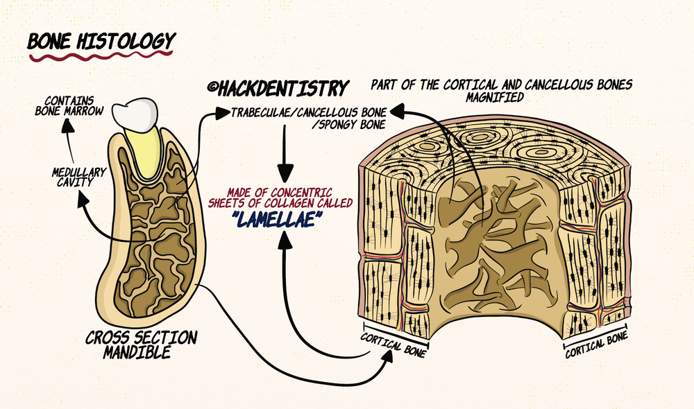
Let’s have a look at the histology of bone with the mandible as an example.
The mandible has a rigid outer shell called the cortical bone or cortex surrounding a medullary cavity or a marrow cavity.
The medullary cavity apart from housing the bone marrow is interrupted by a network of numerous bony spicules or trabeculae called the cancellous bone or the spongy bone.
The cortical and cancellous bones are histologically identical in that, they are made of concentric sheets of collagen called lamellae.
COMPACT BONE HISTOLOGY
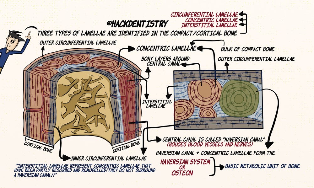
Histologically three types of lamellae can be identified in the compact bone, them being circumferential, concentric and interstitial lamellae.
Bulk of the compact bone is made of concentric lamellae that are actually bony layers wrapped around a central cylindrical tunnel or canal. The central canal is called the Haversian canal and houses blood vessels and nerves.
The Haversian canal and the concentric lamellae together form the Haversian system. The concentric lamellae or the Haversian system form the basic metabolic unit of bone called the osteon.
Circumferential lamellae surround the outer most and the inner most layers of the compact bone.
Lamellae found in between the concentric lamellae are called the interstitial lamellae. The interstitial lamellae actually represent concentric lamellae that have been partly resorbed and remodeled and no longer surround a Haversian canal.
Osteoblasts, Osteocytes and Lacunae
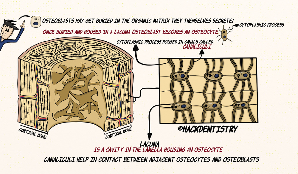
During bone formation when the organic matrix is laid down by osteoblasts, they often get buried within the lamellae and are housed in small cavities called lacunae.
Osteoblasts housed in lacunae are called osteocytes and they have delicate cytoplasmic processes radiating out that are housed in micro canals called canaliculi.
These canaliculi are interconnecting and help osteocytes contact adjacent osteocytes as well as the osteoblasts lining the bone surface.
Periosteum and Nutrition
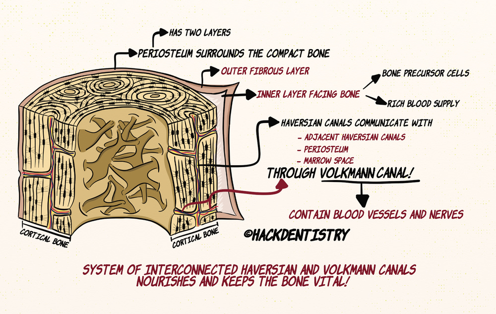
The outer most layer of the compact bone is surrounded by a periosteum having two layers; an outer fibrous layer and an inner layer facing the bone, having precursor bone cells and a rich blood supply.
The Haversian canals communicate with each other as well as the periosteum and the marrow space via canals called Volkmann canals.
Volkmann canals like Haversion canals also contain blood vessels.
This system of interconnected Haversian and Volkmann canals, supplies nutrients and nourishes the bone.
CANCELLOUS/TRABECULAR/SPONGY BONE HISTOLOGY
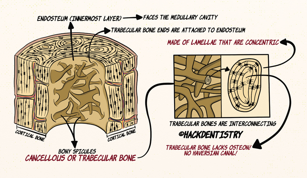
The innermost layer of the compact bone is called the endosteum and is covered by inner circumferential lamellae.
The innermost layer faces the medullary cavity that houses a fine network of bony spicules called the cancellous bone or trabecular bone.
These spicules of trabecular bone are interconnecting and their ends are attached to the endosteum.
Like the compact bone, the trabecular bone is also made of lamellae that are concentric. However, trabecular bone does not have osteons like in the compact bone.
HIGHLIGHTS - VIVA & ENTRANCE EXAM PERSPECTIVE
Osteoblasts, osteocytes and osteoclasts make up the cells of the bone, while the inter-cellular material is made of organic and inorganic material.
Most bones are essentially made of two tissue patterns called
a) compact or cortical bone and a
b) fine network of cancellous bone or spongy/trabecular bone.The cortical and cancellous bones are histologically identical in that, they are made of concentric sheets of collagen called lamellae.
Histologically three types of lamellae can be identified in the compact bone, them being circumferential, concentric and interstitial lamellae.
Bulk of the compact bone is made of concentric lamellae that are actually bony layers wrapped around a central cylindrical tunnel or canal. The central canal is called the Haversian canal and houses blood vessels and nerves.
The Haversian canal and the concentric lamellae together form the Haversian system.
The concentric lamellae or the Haversian system form the basic metabolic unit of bone called the osteon.
Osteoblasts housed in lacunae are called osteocytes and they have delicate cytoplasmic processes radiating out that are housed in micro canals called canaliculi.
The Haversian canals communicate with each other as well as the periosteum and the marrow space via canals called Volkmann canals.
Like the compact bone, the trabecular bone is also made of lamellae that are concentric. However, trabecular bone does not have osteons like in the compact bone.
REFERENCES AND FURTHER READING
Nanci A. Tencate’s Oral Histology. Development, Structure and Function. 8th ed. Elsevier; 2013.
Young B, Woodford P, O’Dowd G. Wheater’s Functional Histology. A Text and Colour Atlas. 6th ed. Elsevier Churchill Livingstone; 2014.
Kumar GS. Orban’s Oral Histology and Embryology.13th ed. Elsevier; 2011.
Lindhe J, Lang NK, Karring T. Clinical periodontology and implant dentistry. 5th ed. Blackwell Munksgaard;2008.