
Desmosomes and Hemi-desmosomes (Note & Video)

👆A part of this video "Oral Mucosa - Epithelium part I" gives an overview of desmosomes.
👆This video "How does the Oral Epithelium attach to the connective tissue" explains how hemidesmosomes play a role in the same.
Epithelial cells are attached to each other and to the connective tissue through specialized structures called “cell junctions“.
There are many different types of cell junctions and are classified as follows:
TYPES OF CELL JUNCTIONS
Tight junctions (also called zonula occludens)
Adhesive junctions
Intercellular / Cell-cell
Zonula adherens
Desmosomes (also called macula adherens)
Cell - matrix
Focal adhesions
Hemidesmosomes
Gap junctions
Table Courtesy - Nanci A. Tencate’s Oral Histology. Development, Structure and Function. 8thed. Elsevier; 2013.
For the sake of brevity, we would only talk about desmosomes and hemi-desmosomes and their clinical relevance in this section.
As you would have noted in the classification, both desmosomes and hemi-desmosomes are adhesive junctions.
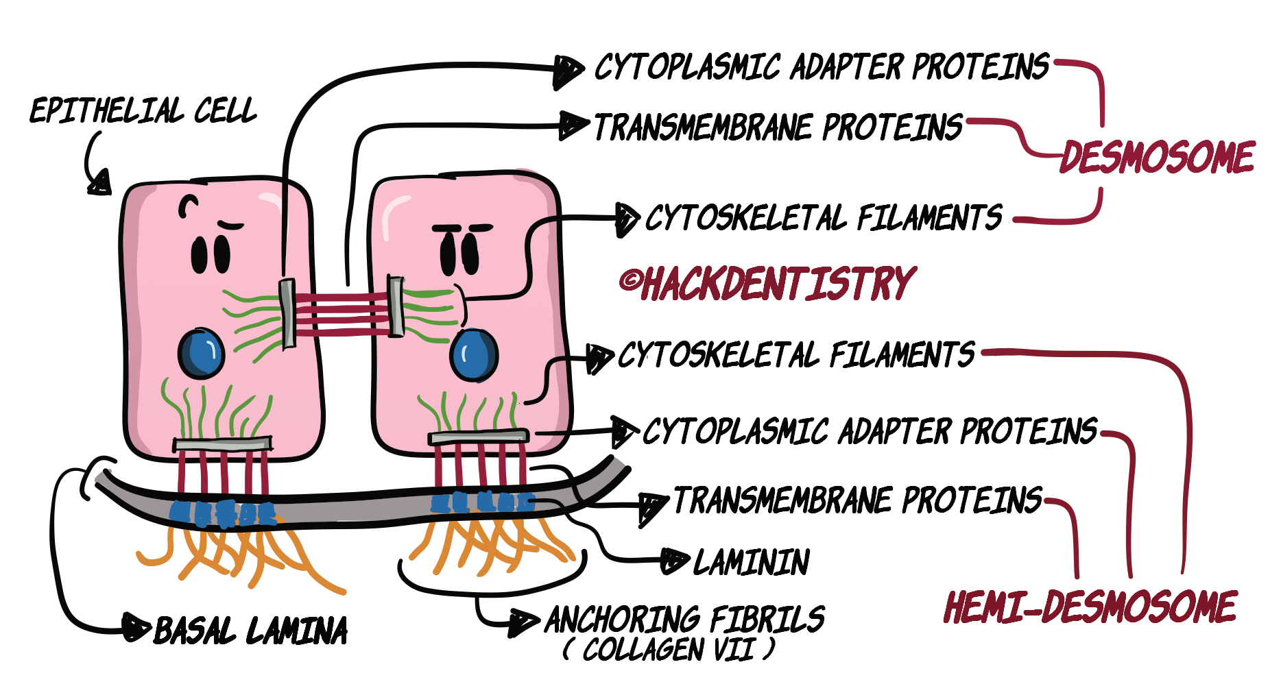
Under the adhesive junctions, note that desmosomes are cell-cell or intercellular junctions and hemidesmosomes are cell - matrix junctions (Figure 2).
💡KEY TAKEAWAYS

- Epithelial cells attach to each other through desmosomes (CELL-CELL).

- Epithelial cells attach to the connective tissue below them, via hemi-desmosomes (CELL - MATRIX).
DESMOSOMES
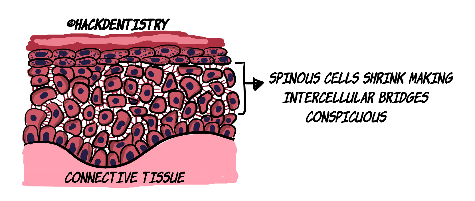
Though desmosomes are ultramicroscopic structures, they can be visualized as “spines”, in light microscopy, in between the spinous cells in the oral epithelium.
During histologic preparations, the epithelial cells (in the spinous layer) shrink away from each other and are in contact only in points where they are attached with each other through desmosomes or “inter-cellular bridges”.
This makes the inter-cellular bridges conspicuous under light microscopy.
Desmosomes are essentially made of three components/proteins:
- Transmembrane proteins
- Cytoplasmic adapter proteins
- Cytoskeletal filaments

(Refer Figure 2)
Transmembrane proteins
As the name explains, the transmembrane protein of an epithelial cell traverses through the cell membrane of the cell to make contact with the transmembrane protein of the adjacent epithelial cell.
Transmembrane proteins in desmosomes are “desmoglein“ and “desmocollin“.
Cytoplasmic adapter proteins
These proteins form dense plaques in the cytoplasm of the cell, that help in attachment of transmembrane proteins and cytoskeletal filaments.
Cytoskeletal filaments
These are intermediate filaments (cytokeratins).
CLINICAL SIGNIFICANCE

- Pemphigus is a skin disease (also having oral manifestations), where the desmoglein proteins (transmembrane proteins) of the desmosomes are primarily targeted by auto-antibodies severing inter-epithelial junctions resulting in a split in the epithelium.

- This results in erosion of the epithelium leading to painful blisters and ulcers both in the skin and oral cavity.
HEMI-DESMOSOMES
Just like how epithelial cells don’t rip apart from each other, the epithelium as a whole, has to firmly sit on the connective tissue without splitting away.
The firm attachment of the epithelium to the connective tissue is enhanced by three factors:
The epithelium-connective tissue interface is undulating or wavy, rather than being flat. This makes for an interface with a larger surface area (than if it was flat) and provides a firmer attachment.
Scanning electron micrographs show that connective tissue has numerous conical papillae jutting out and penetrating the epithelium.
Hemi-desmosomes provide for a firm attachment of the cells with the connective tissue (Figure 2).
A NOTE BASEMENT MEMBRANE/BASAL LAMINA
Understand that epithelial cells do not directly sit on the connective tissue per se.
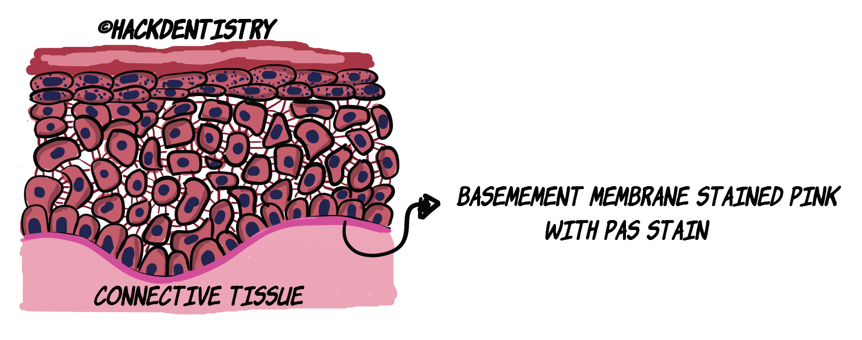
The epithelial cells are separated from the connective tissue by a “basement membrane“.
In the light microscope, when the tissue is stained with a special stain called Periodic Acid Schiff (PAS), the basement membrane can be visualized as a thin (pink) line between the epithelium and the connective tissue.
However, at the ultrastructural level, the basement membrane is called basal lamina and consists of 3 zones called -> a) lamina lucida, b) lamina densa and c) lamina fibroreticularis.
Like desmosomes, hemi-desmosomes also comprise of three proteins:

- Transmembrane proteins
- Cytoplasmic adapter proteins
- Cytoskeletal filaments

Refer Figure 2
Transmembrane proteins
In the hemidesmosomal junction Bullous Pemphigoid antigen 180 (BP 180) and α6β4 integrins form the transmembrane proteins.
Cytoplasmic adapter proteins
These proteins form dense plaques in the cytoplasm of the cell, that help in attachment of transmembrane proteins and cytoskeletal filaments. BP 230 and plectin form the cytoplasmic adapter proteins.
Cytoskeletal filaments
These are intermediate filaments (cytokeratins).
Together, these proteins along with other constituents of the basal lamina, like laminin and type VII collagen otherwise called the anchoring fibrils, help in firm attachment of the epithelium to the connective tissue.
CLINICAL SIGNIFICANCE

- Mucous membrane pemphigoid, is an auto-immune disease characterized by formations of blisters and ulcers in the oral cavity and skin.

- In this disease auto-antibodies attack components of the basement membrane zone (hemidesmosomal junction), predominantly bullous pemphigoid antigen 180 (BP180). causing epithelium to detach from the connective tissue, leading to the formation of blisters and ulcers.

- Other components of the basement membrane that may be attacked (less often) are BPA2/BP230, laminin5, type VII collagen and β4 subunit of α6β4 integrin.
HIGHLIGHTS - VIVA & ENTRANCE EXAM PERSPECTIVE
Epithelial cells attach to each other through desmosomes (CELL-CELL junctions).
Epithelial cells attach to the connective tissue below them, via hemi-desmosomes (CELL - MATRIX junctions).
During histologic preparations, the epithelial cells (in the spinous layer) shrink away from each other and are in contact only in points where they are attached with each other through desmosomes or “inter-cellular bridges”.
Desmosomes and hemi-desmosomes are essentially made of three components/proteins -> a) Transmembrane proteins, b) Cytoplasmic adapter proteins, and c) Cytoskeletal filaments.
Transmembrane proteins in desmosomes are “desmoglein“ and “desmocollin“.
Pemphigus is a skin disease (also having oral manifestations), where the desmoglein proteins (transmembrane proteins) of the desmosomes are primarily targeted by auto-antibodies severing inter-epithelial junctions resulting in a split in the epithelium.
Hemi-desmosomes provide for a firm attachment of the cells with the connective tissue.
In the light microscope, when the tissue is stained with a special stain called Periodic Acid Schiff, the basement membrane can be visualized as a thin (pink) line between the epithelium and the connective tissue.
However, at the ultrastructural level, the basement membrane is called basal lamina and consists of 3 zones called -> a) lamina lucida, b) lamina densa and c) lamina fibroreticularis.
The basal lamina consists of proteins making up hemi-desmosomes.
In the hemidesmosomal junction Bullous Pemphigoid antigen 180 (BP 180) and α6β4 integrins form the transmembrane proteins.
In mucous membrane pemphigoid auto-antibodies attack components of the basement membrane zone (hemidesmosomal junction), predominantly bullous pemphigoid antigen 180 (BP180).
REFERENCES AND FURTHER READING
Berkovitz BKB, Hollan GR, Moxham BJ. Oral Anatomy, Histology and Embryology. 4th ed. Mosby Elsevier; 2009.
Nanci A. Tencate’s Oral Histology. Development, Structure and Function. 8th ed. Elsevier; 2013.
Kumar GS. Orban’s Oral Histology and Embryology.13th ed. Elsevier; 2011.
Avery JK. Oral development and Histology. 3rd ed. Thieme Medical Publishers; 2002.