
Development and growth of teeth Part II: Bell stage and root formation (Note & Video)

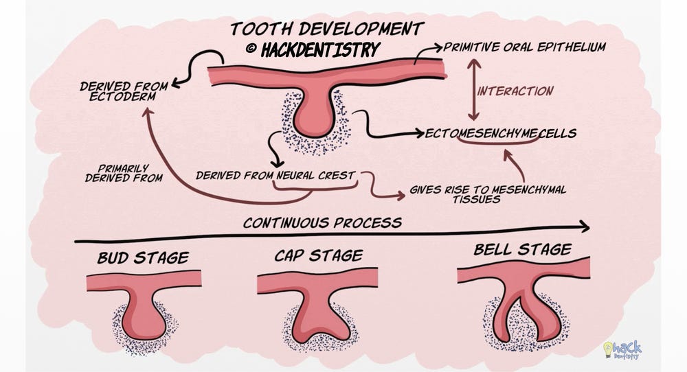
The development and growth of teeth is a complex process of interactions between the primitive oral epithelium and the underlying ectomesenchymal cells.
The epithelium is derived from the ectoderm of the first pharyngeal arch while the cells of the ectomesenchyme are neural crest in origin.
Since the neural crest cells are primarily derived from the ectoderm and eventually give rise to mesenchymal tissues, they are called ectomesenchymal cells.
Tooth development, though a continuous process, can be divided into three stages called the bud, cap and bell stages. These stages are so named because of the shape the enamel organ assumes in each stage.
BELL STAGE
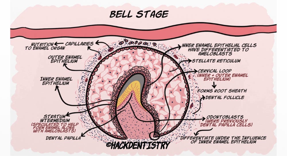
In this stage, the enamel organ continues to grow and assumes a bell shape. Before we get into details of the events happening in the bell stage, let’s have a look at the different types of cells constituting the whole tooth germ at this stage.
Like the cap stage, the early bell stage comprises of an inner enamel epithelium lining the concavity of the bell and an outer enamel epithelium lining the periphery of the enamel organ. There are capillary plexuses established near the outer enamel epithelium which bring in nutrition for the cells of the enamel organ. The inner enamel epithelial cells differentiate in this stage to become tall columnar ameloblasts!
Some cells between the inner enamel epithelium and stellate reticulum become spindle shaped and form a layer called stratum intermedium. These cells are speculated to work in tandem with the inner enamel epithelium to form enamel.
The rim of the enamel organ at the cervical region where the inner enamel epithelium meets the outer enamel epithelium is called the cervical loop. The cervical loop is an important part of the tooth germ in that; it gives rise to the root sheath, which proliferates to form the root.
The cells below the concavity are called dental papilla and those surrounding the enamel organ and the dental papilla constitute the dental follicle. The dental papilla cells near the inner enamel epithelium differentiate under their influence to become odontoblasts.
Events in Bell Stage
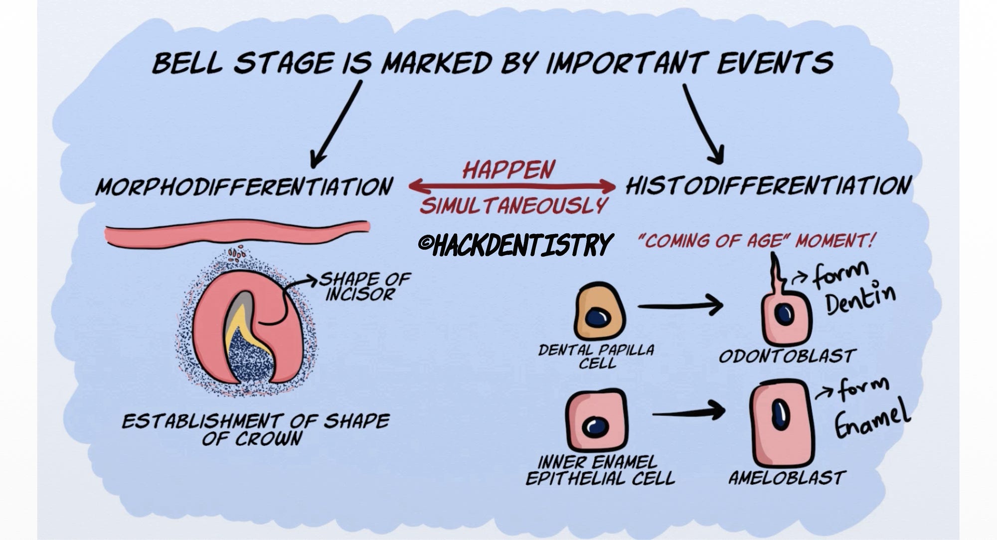
The bell stage is marked by two events called morphodifferentiation and histodifferentiation, both happening simultaneously.
Morphodifferentiation refers to the establishment of the shape of the crown.
Histodifferentiation refers to the “coming of age” moment, when the dental papilla cells differentiate to become odontoblasts and the inner enamel epithelial cells become ameloblasts. Odontoblasts form dentin and ameloblasts form enamel.
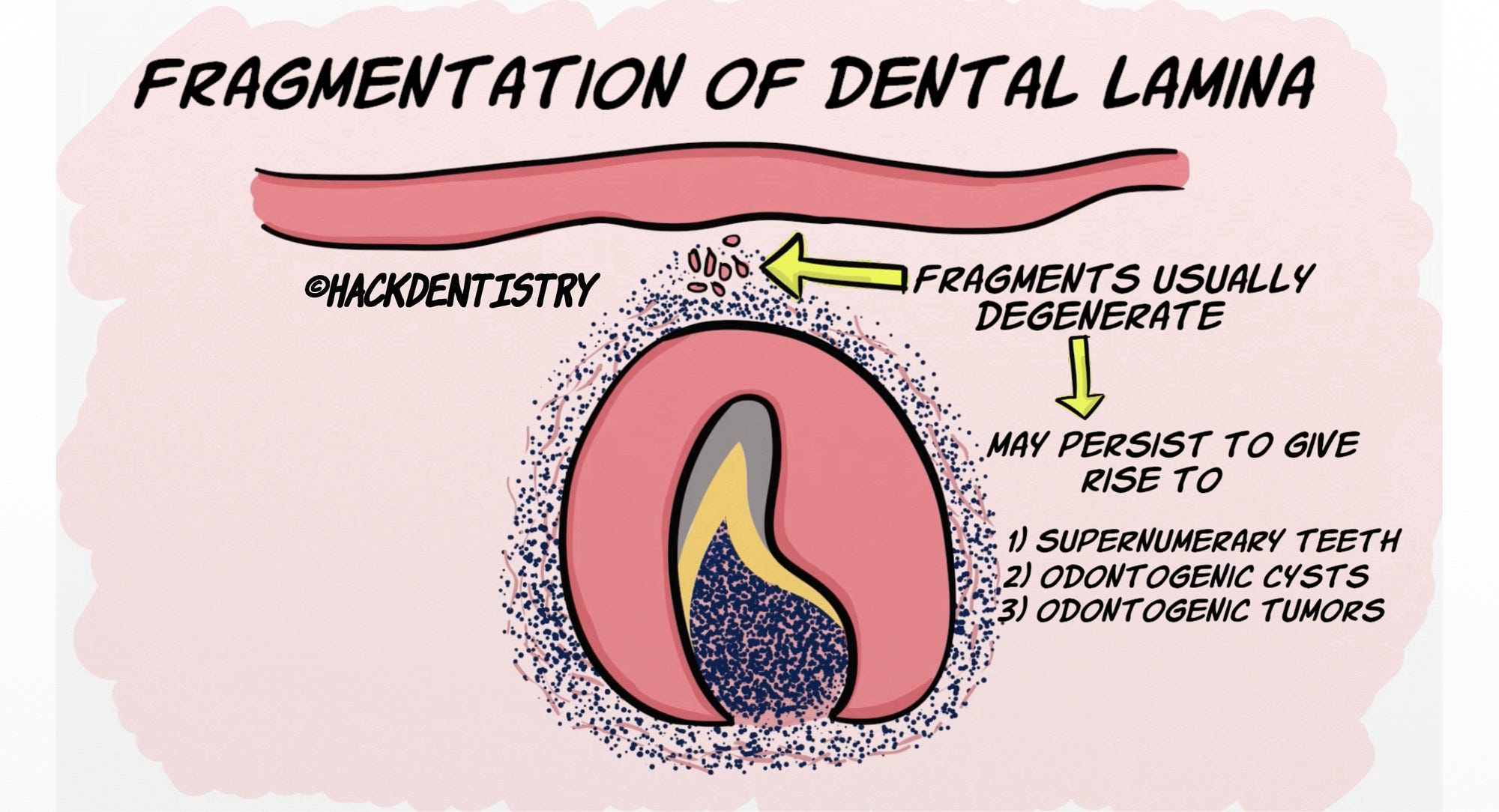
Another important event happening in the bell stage is the fragmentation of the dental lamina. The dental lamina disintegrates, separating the tooth germ from the oral epithelium. These fragments of the dental lamina degenerate.
However, they may sometimes persist giving rise to supernumerary teeth, odontogenic cysts and tumors.
Acquiring a “Bell” shape
It was initially thought that the ectomesenchyme exerted pressure on the inner enamel epithelium, giving it the bell shape thereby determining the shape of the crown.
However, that is not the case. Just like the cap stage, the enamel organ assumes a bell shape because of differential growth of cells.
Let’s try to understand how this happens and remember that morpho- and histo-differentiation happen simultaneously.
The inner enamel epithelium, in the early bell stage secrete growth factors and signalling molecules which help in differentiation of dental papilla cells to odontoblasts.
Simultaneously, the inner enamel epithelial cells corresponding to the future cusp tip position begin to differentiate into ameloblasts. Once differentiated, they stop proliferating!
However the adjacent inner enamel epithelial cells that have not differentiated continue to divide, exerting a pressure. Also, the inner enamel epithelium is constrained and restricted by the cervical loop.
So, the pressure exerted by the dividing cells and the confinement of these cells by the cervical loop results in these cells buckling and assuming the shape of the cusp of the corresponding tooth being formed.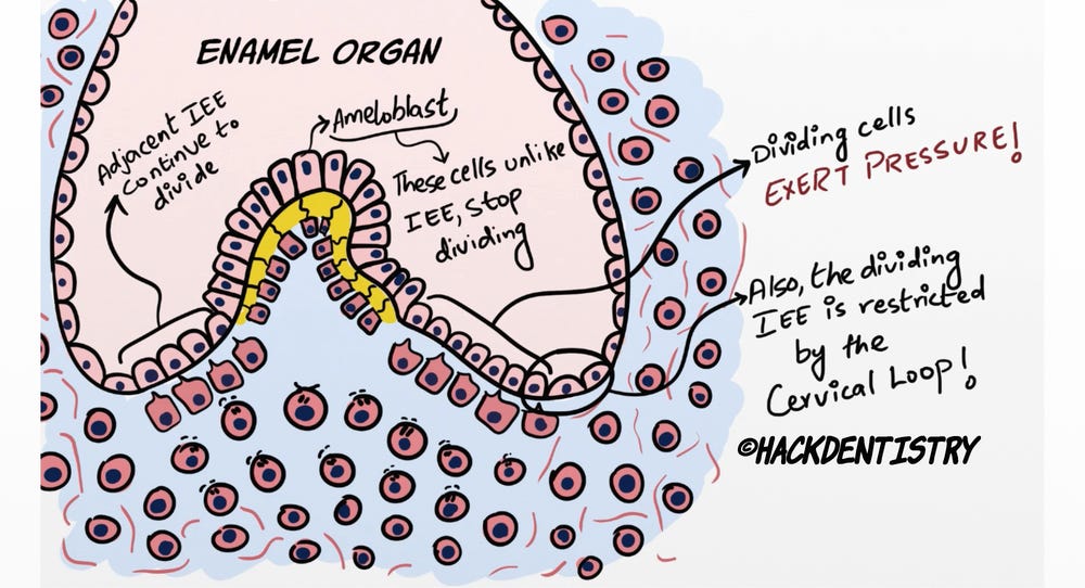
NOTE

The inner enamel epithelium and the dental papilla continue to differentiate from the cusp tip down the slopes of the cusp to the cervical region. Hence the deposition of dentin and enamel begins at the cusp tip and ends cervically.
Advanced Bell Stage
The advanced bell stage is marked by the formation and mineralization of the enamel and dentin.
When enamel and dentin form incrementally, the stellate reticulum collapses.
Finally the ameloblasts and the outer enamel epithelium fuse with each other, forming the reduced enamel epithelium surrounding the entire crown!
ROOT FORMATION
Once the crown is formed, the rim of the enamel organ at the cervical portion comprises the inner and outer enamel epithelium and is called the cervical loop. This cervical loop starts to proliferate to give rise to the Hertwig’s epithelial root sheath.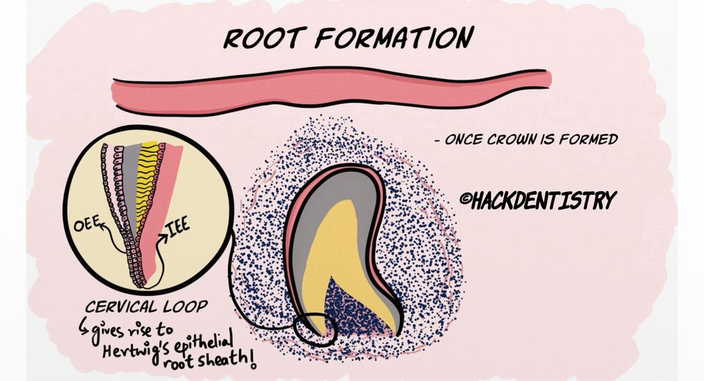
Root Sheath and Epithelial Diaphragm
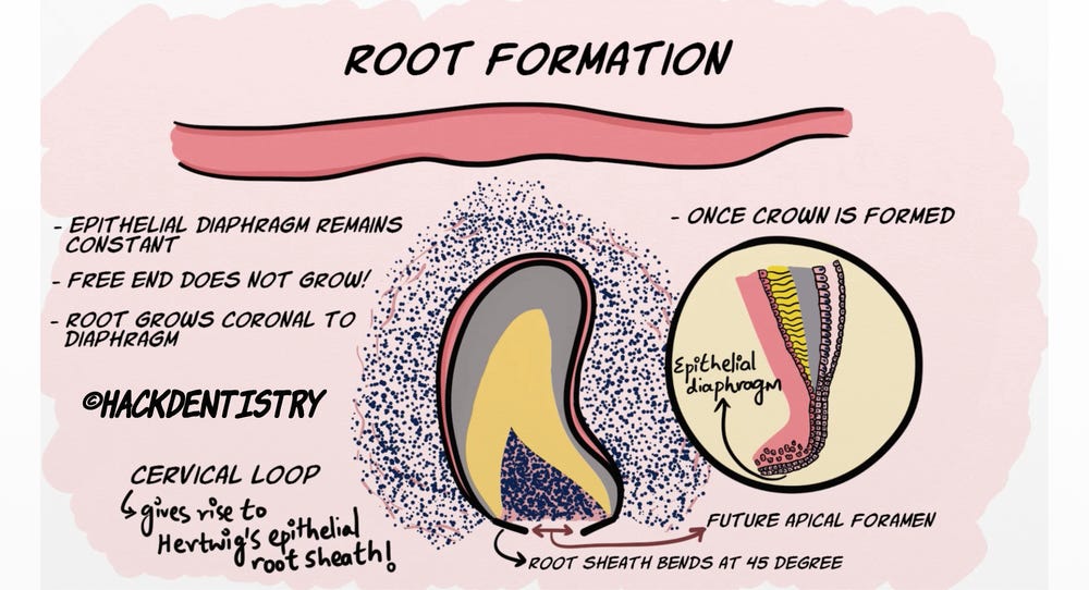
At the beginning the root sheath bends at an angle of 45 degrees towards the pulp constricting or narrowing the wide cervical opening of the crown.
This is called the epithelial diaphragm and the opening would eventually become the apical foramen.
The epithelial diaphragm remains constant and the free end does not grow. What rather happens is that the root sheath grows coronal to the diaphragm.
Rest of Root Formation
As the root sheath proliferates, the inner layer of the root sheath induces the differentiation of the adjacent dental papilla cells to odontoblasts and these odontoblasts form dentin.
As dentin is formed and is mineralizing, the adjacent root sheath cells start to disintegrate.
Now, it has to be understood that the root sheath is never continuous. It keeps disintegrating as it grows.
Most of the disintegrated cells move away from the root surface, and cells from the dental follicle interact with the dentin.
On interaction these cells differentiate to form the cementoblasts and lay down cementum.
Some of the disintegrated cells do not migrate and may persist in the root area. These cells form the epithelial rests of Malassez.Root formation in multi-rooted teeth
As for multi-rooted teeth, the root sheath proliferation takes place in the same manner as single rooted teeth.
However, the diaphragm develops tongue like extensions two or three in number depending on the tooth being formed.
These extensions fuse with each other dividing the opening into two or three openings.
REFERENCES AND FURTHER READING
Avery JK.Oral development and Histology.3rd ed. Thieme Medical Publishers;2002.
Nanci A. Tencate’s Oral Histology. Development, Structure and Function. 8th ed. Elsevier;2013.
Kumar GS. Orban’s Oral Histology and Embryology.13th ed. Elsevier;2011.