
Structural features of enamel (Note)

In this note we would be discussing important structural features of enamel, them being:
Enamel rods
Striae or Incremental lines of Retzius
Enamel tufts
Enamel spindles
Enamel lamellae
Hunter Schreger bands
ENAMEL RODS
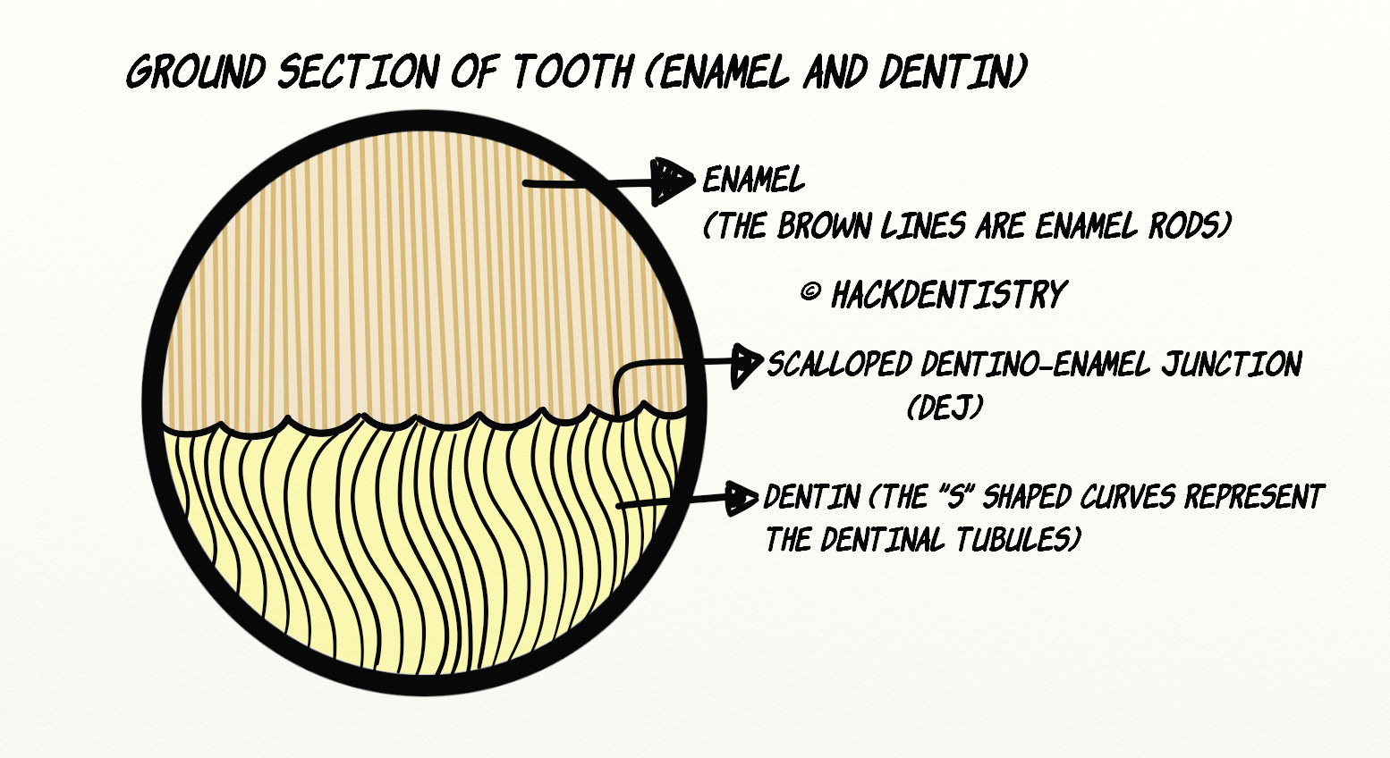
Enamel rod also called enamel prism forms the basic structural unit of enamel.
Enamel rods consist of densely packed crystallites and run from the DEJ to the enamel.
Enamel rods have an undulating or tortuous course and therefore it’s length is greater than the thickness of the enamel.
PATTERNS OF ROD/PRISM ARRANGEMENT
Pattern I
Consists of circular enamel rods surrounded by interrod or inter-prismatic enamel.

Pattern II
Has a stacked or a vertical pattern.

Pattern III
In this pattern the enamel rods are arranged in such a way that they have a “keyhole” appearance.
The convex surface of the rod is called the “head” and is oriented towards the cusp or incisal region. The neck region of the “keyhole” is called the “tail” of the enamel rod and is oriented apically.

Note

Some investigators are of the view that the “tail” region of the enamel rod actually represents the interred or interprismatic substance.
BASICS OF GROUND SECTION OF TOOTH (ENAMEL AND DENTIN)
STRIAE OF RETZIUS
Striae of Retzius also called incremental lines of Retzius appear as brownish lines running from the DEJ across the enamel obliquely. It appears this way in ground sections that are cut longitudinally.
However in transverse/cross sections, Striae of Retzius appear as concentric rings.
Striae of Retzius are ascribed to be formed due to incremental or appositional growth of enamel. They represent the enamel forming front.
Incremental lines/Striae of Retzius (Longitudinal section)

Incremental lines/Striae of Retzius (Transverse section)

ENAMEL TUFTS
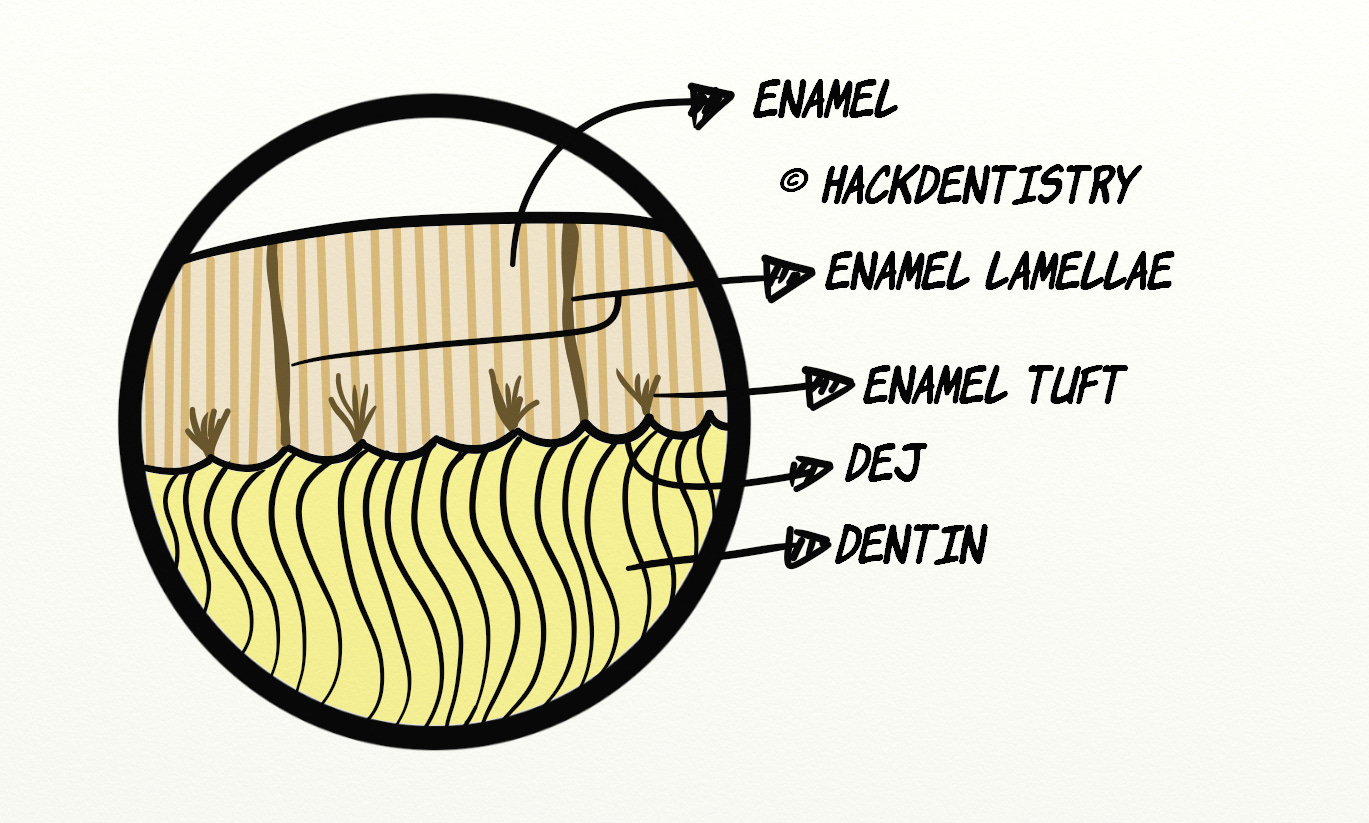
Enamel tufts are hypomineralized structures with a high concentration of organic content (protein). They contain enamel rods that are hypocalcified.
The inner end of enamel tufts project from the DEJ and extend to a short distance in the enamel (about 1/3rd the enamel thickness).
This structure is so named because it resembles a “tuft of grass” when viewed at low magnification.
Enamel tufts are best seen in transverse or cross sections of enamel.
The protein content of enamel is highest in the enamel tuft. It contains “tuftelin” a non-amelogenin group of proteins.
ENAMEL SPINDLES
Enamel spindles are narrow tubule like hypomineralized structures arising from the DEJ and extending up to 25 microns into the enamel.
It is thought that, during the early stage of amelogenesis, some odontoblast processes may project beyond the inner enamel epithelial region by inserting themselves between these cells (inner enamel epithelium or pre-ameloblasts).
It is also thought to be remnants of dead odontoblasts.
Enamel spindles are numerous below the incisal and cuspal regions.
Enamel spindles are better seen in longitudinal sections.
Enamel spindles appear dark in transmitted light.

ENAMEL LAMELLAE
Enamel lamellae are sheet like structural faults, running across the full thickness of the enamel up to the DEJ.
These structures are narrow, hypomineralized and filled with organic material.
It is known to develop due to incomplete maturation of enamel rods.
There may be 3 types of lamellae:

Type A: lamellae made of poorly calcified segments of rods
Type B: lamellae composed of degenerated cells
Type C: lamellae filled with organic matter which may originate from saliva
Lamellae may be a gateway for entry of bacteria that may cause caries. It is therefore a site of weakness.

Note

What appears to be enamel lamellae in the ground section could actually be cracks that may occur during preparation of the ground section. Enamel lamellae and “cracks” appear to be similar when viewed under the microscope. Lamellae can be however distinguished from cracks by demineralization. On doing so, cracks would disappear while lamellae would not.
HUNTER-SCHREGER BANDS
Hunter-Schreger bands are produced as a result of an optical phenomenon that occurs due to changes in the direction of enamel rods.
These bands appear as alternating light and dark bands under reflected light and can be seen in longitudinal sections.
The light zones represent longitudinally sectioned rods, known as parazones and the dark bands correspond to cross-sectional enamel rods, known as diazones.
HIGHLIGHTS - VIVA & ENTRANCE EXAM PERSPECTIVE
Enamel rod also called enamel prism forms the basic structural unit of enamel.
Patterns of rod/prism arrangement:
Pattern I -> Consists of circular enamel rods surrounded by interrod or inter-prismatic enamel.
Pattern II -> Has a stacked or a vertical pattern
Pattern III -> a) In this pattern the enamel rods are arranged in such a way that they have a “keyhole” appearance. b)The convex surface of the rod is called the “head” and is oriented towards the cusp or incisal region. c) The neck region of the “keyhole” is called the “tail” of the enamel rod and is oriented apically.
Striae of Retzius appear as brownish lines running from the DEJ across the enamel obliquely.
Striae of Retzius are ascribed to be formed due to incremental or appositional growth of enamel. They represent the enamel forming front.
Enamel tufts are hypomineralized structures with a high concentration of organic content (protein). They contain enamel rods that are hypocalcified.
Enamel tuft is so named because it resembles a “tuft of grass” when viewed at low magnification.
Enamel tufts are best seen in horizontal or cross sections of enamel.
Enamel spindles are narrow tubule like hypomineralized structures arising from the DEJ and extending up to 25 microns into the enamel.
Enamel spindles are numerous below the incisal and cuspal regions.
Enamel spindles are better seen in longitudinal sections.
Enamel lamellae are sheet like structural faults, running across the full thickness of the enamel up to the DEJ.
There may be 3 types of lamellae:
Type A: lamellae made of poorly calcified segments of rods
Type B: lamellae composed of degenerated cells
Type C: lamellae filled with organic matter which may originate from saliva
Lamellae may be a gateway for entry of bacteria that may cause caries. It is therefore a site of weakness.
Lamellae can be distinguished from “cracks” by demineralization. On doing so, cracks would disappear while lamellae would not.
These bands appear as alternating light and dark bands under reflected light and can be seen in longitudinal sections.
The light zones represent longitudinally sectioned rods, known as parazones and the dark bands correspond to cross-sectional enamel rods, known as diazones.
REFERENCES AND FURTHER READING
Berkovitz BKB, Hollan GR, Moxham BJ. Oral Anatomy, Histology and Embryology. 4th ed. Mosby Elsevier; 2009.
Nanci A. Tencate’s Oral Histology. Development, Structure and Function. 8th ed. Elsevier; 2013.
Kumar GS. Orban’s Oral Histology and Embryology.13th ed. Elsevier; 2011.
Avery JK. Oral development and Histology. 3rd ed. Thieme Medical Publishers; 2002.