
Erythroplakia (Notes & Video)

“Erythro” stands for red and “plakia” for patch. Erythroplakia is a red patch occurring in the oral mucosa, which cannot be diagnosed as any other definable lesion.
Erythroplakia defined as --> “A fiery red patch that cannot be characterized clinically or pathologically as any other definable disease”.
💡Know Thy Facts!
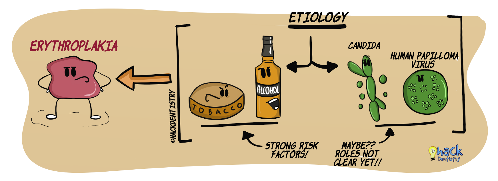
Erythroplakia is a diagnosis of exclusion. There are a number of red lesions in the oral cavity resembling erythroplakia. These lesions are clinico-histopathologically definable lesions. For example, oral lichen planus (OLP) may manifest as an atrophic or erosive red lesion resembling erythroplakia. However, OLP has definitive clinical (white striae elsewhere in the oral cavity) and histopathology features. A clinician should rule out such definitive red lesions, before a clinical diagnosis of “erythroplakia” can be made.Tobacco and alcohol are suggested to be important etiologic factors in causing erythroplakia.
Candida albicans has been demonstrated in erythroplakia and erythro-leukoplakia cases. However its role is still unclear.
HPV is suspected to be an etiologic co-factor.
CLINICAL FEATURES
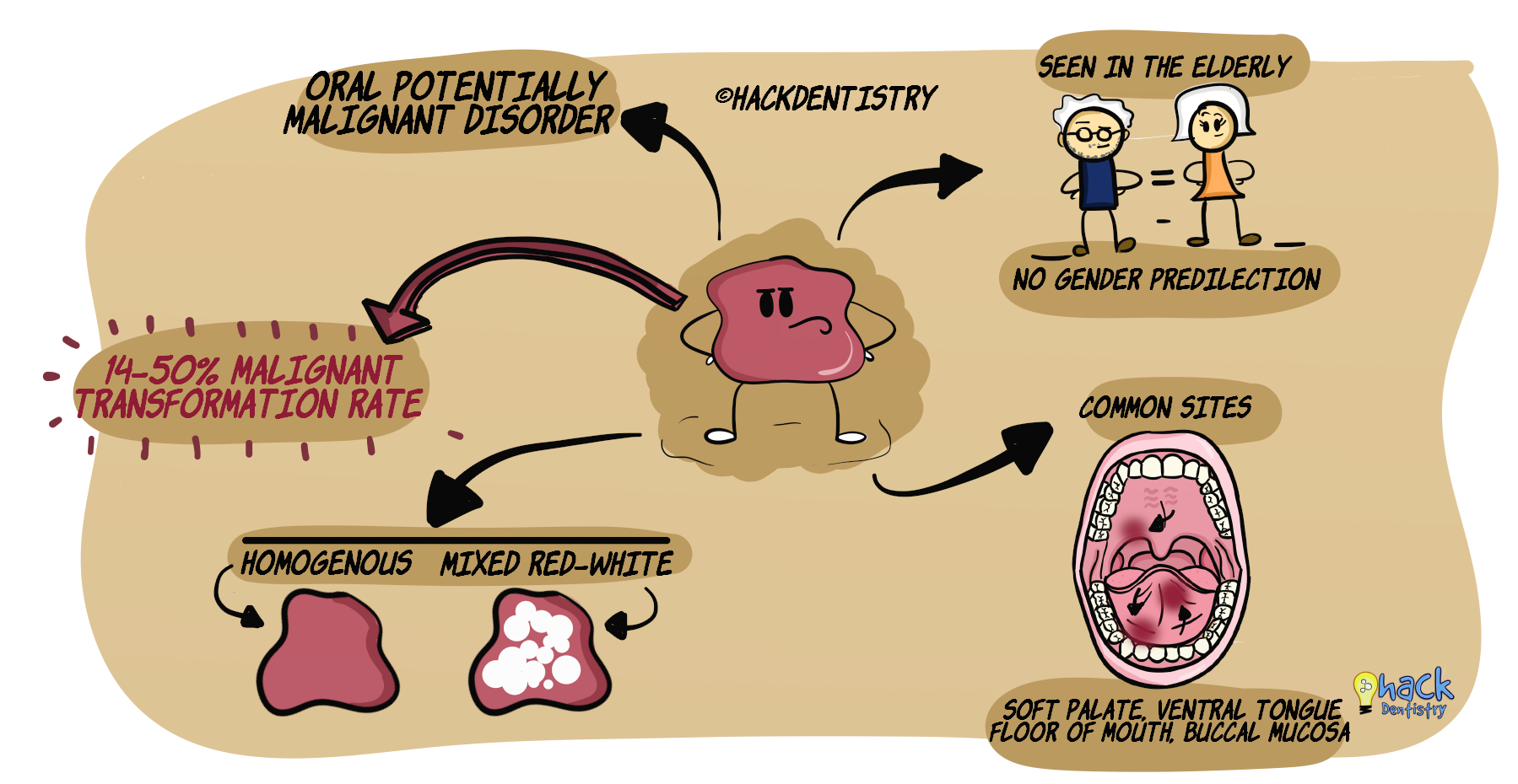
Erythroplakia is a rare potentially malignant disorder and has a speculated worldwide prevalence of 0.02 - 0.83%.
Manifests in the elderly (45-75 years); no gender predilection.
Erythroplakia may occur in any site in the oral cavity; common sites --> soft palate, floor of mouth, buccal mucosa, and ventral tongue.
Erythroplakia may be soft, velvety and red with or without regular margins.
Erythro-leukplakia --> granular specks of white foci interspersed in the erythroplakia lesion. However, there is no significance in denoting a lesion as speckled leukoplakia, leuko-erythroplakia or erythro-leukoplakia. All mixed red-white lesions have to be treated with caution as they have an increased risk of malignancy.
Erythroplakia may manifest adjacent other lesions like leukoplakia, squamous cell carcinoma and oral lichen planus.
Erythroplakia has a high malignant transformation rate, estimated to range from 14-50%.
💡Know Thy Facts!
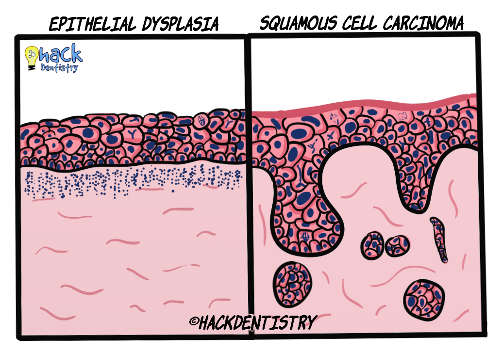
Erythroplakia should be a bigger source of worry to the clinician than leukoplakia. Most erythroplakia lesions show dysplasia, carcinoma in-situ or squamous cell carcinoma and have a higher rate of malignant transformation than leukoplakia. HISTOPATHOLOGY FEATURES
Findings for most cases of erythroplakia range from --> dysplasia (mild/moderate/severe) --> carcinoma-in situ --> squamous cell carcinoma (refer topic on “Epithelial dysplasia”).
TREATMENT
Since most erythroplakia cases show dysplasia and invasive carcinoma, a complete excision is the treatment of choice.
Long term follow up of patients after treatment.
✅HIGHLIGHTS - VIVA & ENTRANCE EXAM PERSPECTIVE
Erythroplakia is a fiery red patch that cannot be characterized clinically or pathologically as any other definable disease.
Erythroplakia is a rare potentially malignant disorder.
Apart from erythroplakia , mixed red-white lesions also have to be treated with caution as they have an increased risk of malignancy.
Erythroplakia has a high malignant transformation rate, estimated to range from 14-50%.
Erythroplakia should be a bigger source of worry to the clinician than leukoplakia. Most erythroplakia lesions show dysplasia, carcinoma in-situ or squamous cell carcinoma and have a higher rate of malignant transformation than leukoplakia.
📖REFERENCES AND FURTHER READING
Reichart PA, Philipsen HP. Oral erythroplakia--a review. Oral Oncol. 2005 Jul;41(6):551-61.
Villa A, Villa C, Abati S. Oral cancer and oral erythroplakia: an update and implication for clinicians. Aust Dent J. 2011 Sep;56(3):253-6.
Warnakulasuriya S. Clinical features and presentation of oral potentially malignant disorders. Oral Surg Oral Med Oral Pathol Oral Radiol. 2018 Jun;125(6):582-590.
van der Waal I. Potentially malignant disorders of the oral and oropharyngeal mucosa; terminology, classification and present concepts of management. Oral Oncol. 2009 Apr-May;45(4-5):317-23.
Neville BW, Damm DD, Allen CM, Chi A. Oral and Maxillofacial Pathology. South Asian ed. Elsevier; 2016.