
Keratinized and Non-keratinized epithelium (Note & Video)

The oral mucosa is lined by a stratified squamous epithelium.
The epithelium may follow two patterns of maturation, maturing to form either the keratinized or the non-keratinized epithelium.
Except for the gingiva, hard palate and dorsal tongue (which is lined by the keratinized epithelium) the rest of the oral mucosa is lined by the non-keratinized epithelium.
The epithelium is cuboidal at the lower most layer and gradually becomes larger and flatter as it moves up.
The lower most layer of the epithelium, comprising of the basal cells have stem cells which divide to produce daughter cells that differentiate and mature as they move up layers.
NOTE

Even though mucosa over the tongue is designated as "specialized mucosa", the epithelium is actually keratinized. The mucosa is so designated due to the presence of different types of papillae and taste buds.
A BRIEF NOTE ON CELL JUNCTIONS
The cells of the oral epithelium firmly attach to each other with the help of “intercellular junctions“.
These intercellular junctions referred to as “intercellular bridges” (in light microscopy) are called “desmosomes“ or “macula adherens“.
The basal cells attach to the connective tissue below, with the help of “cell-to-matrix junctions“ called “hemi-desmosomes“ or “zonula adherens“.
For now, know that both these junctions consist of three components -> a) Transmembrane proteins, b) Cytoplasmic adapter proteins, c) Cytoskeletal filaments.
LAYERS OF KERATINIZED EPITHELIUM
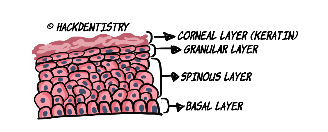
The keratinized epithelium consists of 4 layers or strata namely
the basal layer or stratum basale,
the prickle cell/spinous layer or stratum spinosum,
the granular layer or stratum granulosum and
the corneal/keratinized layer or stratum corneum
💡 KNOW THY FACTS

Epithelial cells are also called keratinocytes. This is due to the presence of tonofilaments, that are keratin intermediate filaments (cytokeratins).
Basal layer or Stratum basale
Light Microscopy
This is a single layer of cells, present just above (adjacent to) the connective tissue (lamina propria).
The basement membrane or basal lamina separates the basal layer from the connective tissue.
The basal cells are cuboidal or low columnar in shape.
This layer houses the stem cells, which divide (mitotic activity) to produce daughter cells for maturation.
In fact, routine staining, could sometimes reveal mitotic figures in this layer.
Ultramicroscopy
Most importantly, basal cells help the epithelium adhere to the connective tissue through hemi-desmosomal junctions.
Without these junctions, the epithelium would rip apart from the connective tissue.
Prickle cell/Spinous layer or Stratum spinosum
Light Microscopy
Above the basal layer is a thick layer of cells called the spinous layer.
The spinous layer forms the bulk of the epithelium.
Spinous cells are spherical to polyhedral in shape.
The single layer of stratum spinosum immediately above the basal layer is called the “parabasal layer“.
Mitotic figures can occasionally be seen in the parabasal layer.
The basal and the parabasal layer are together referred to as “stratum germinativum“.
💡 KNOW THY FACTS
- During histologic preparations, the epithelial cells shrink away from each other and are in contact only in points where they are attached with each other through desmosomes or “inter-cellular bridges” .

- This makes the inter-cellular bridges conspicuous under light microscopy and they appear like “spines”.

Hence the name of the layer -> “spinous or prickle cell“.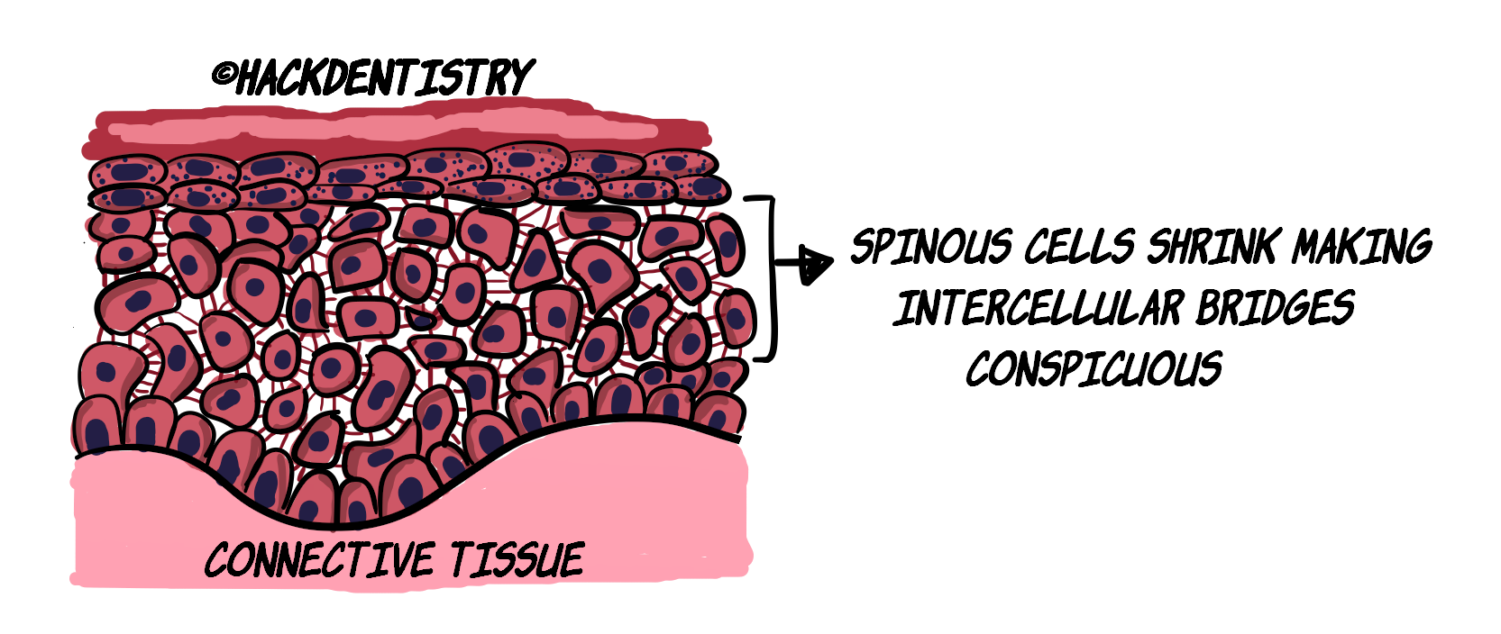
Ultramicroscopy
This layer has conspicuous tonofibrils.
The upper layers of the spinous layer have an organelle called “membrane-coating granule“. It is also called lamellate granule or Odland body.
Membrane coating granules consist of phospholipids.
💡 KNOW THY FACTS

Tonofilaments are keratin intermediate filaments present in epithelial cells.
Tonofilaments, in the keratinized epithelium bundle to form tonofibrils.
In the non-keratinized epithelium, however, tonofilaments are dispersed and are less conspicuous.
Granular layer or Stratum granulosum
Light Microscopy
The granular layer is 3-5 cells thick and comprises of larger and flat cells.
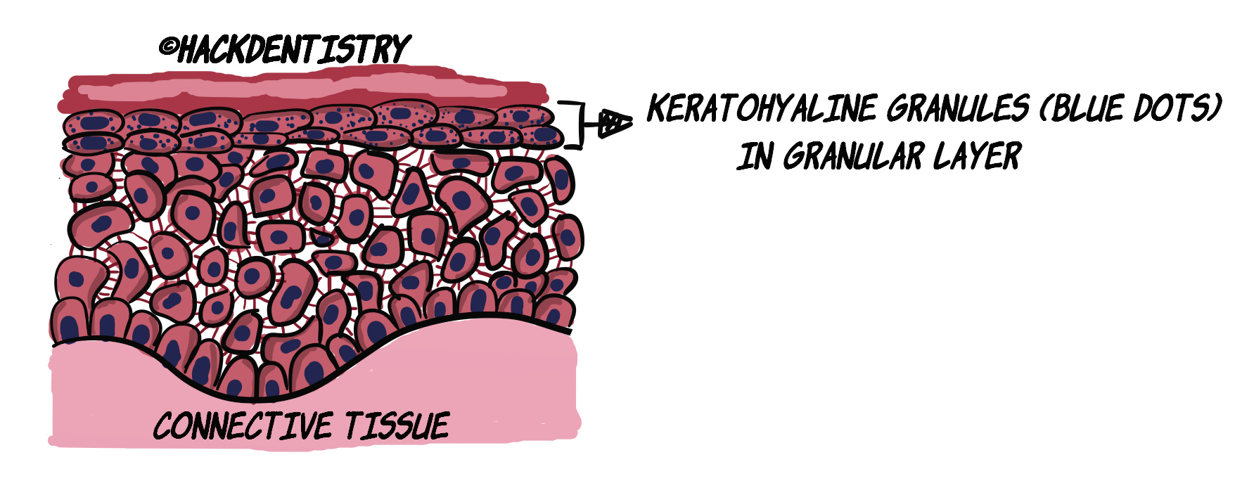
The granular cell is characterized by the presence of keratohyaline granules (hence the name for this layer).
These granules are basophilic (appear as blue dots in under the light microscopy).
Ultramicroscopy (watch the video [at 2:04] to visualize this and understand better)
The granular layer also houses membrane coating granules.
Superficially in the granular layer, the membrane coating granules discharge their content into the intercellular space.
This forms an effective lipid barrier against aqueous substances.
Also evident in the granular layer is the presence of a “cornified cell envelope“.
This envelope lines the inner aspect of the cell membrane of the granular cell.
💡 KNOW THY FACTS

- A protein called "involucrin" forms the main constituent of the cornified cell envelope.

- Keratohyaline granules contain proteins called "filaggrin" and "loricrin".
Corneal/Keratinized layer or Stratum corneum
Light Microscopy
The cells in this layer are dehydrated, have lost all their organelles including their nuclei.
These cells appear like flat disques and are termed “epithelial squames”.
Cells in this layer are constantly shed to be replaced by cells from the layers below.
They appear bright pink under the microscope.
It is a thick layer, providing for mechanical protective function to the masticatory mucosa.
Ultramicroscopy
The corneal cells are densely packed with tonofibrils in matrix of filaggrin, strongly cross-linked with disulphide bonds. This helps in offering resistance and protection to the mucosa.
This mix of tonofibrils in a matrix of filaggrin is collectively called keratin.
💡 KNOW THY FACTS

- The corneal layer in the keratinized epithelium are usually devoid of nuclei. This pattern of keratinization is called "orthokeratinization".

- There are parts of the masticatory mucosa, where the corneal layer retains some pyknotic nuclei. This pattern is referred to as "parakeratinization".
LAYERS OF NON-KERATINIZED EPITHELIUM
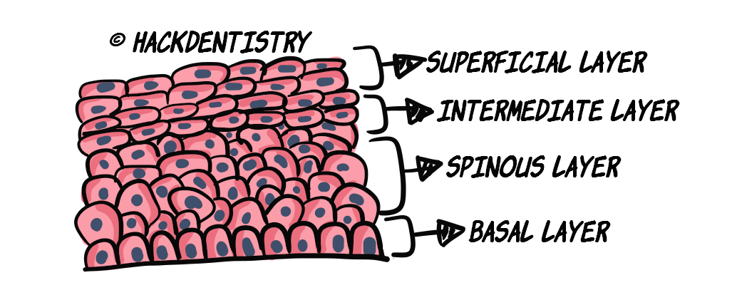
The non-keratinized epithelium consists of 4 (or 3) layers or strata namely
the basal layer or stratum basale,
the prickle cell/spinous layer or stratum spinosum,
the intermediate layer or stratum intermedium and
the superficial layer or stratum superficiale
Some prefer to avoid the use of the term prickle cell or spinous layer and consider only 3 layers in the non-keratinized epithelium.
The first two layers in the non-keratinized epithelium are the same as the keratinized epithelium -> basal and spinous layers.
Non-keratinized epithelium does not have the granular and the corneal layer. Instead the outer two layers are called intermediate layer or stratum intermedium and superficial layer or stratum superficiale.
Non-keratinized epithelium is so designated due to the absence of the corneal or keratinized layer.
Non-keratinized epithelium lacks keratohyaline granules.
The cells in the stratum intermedium have membrane coating granules. But they differ in being round as opposed to the ovoid granules in the keratinized epithelium.
These granules also secrete lipid material into the intercellular space between the intermediate and superficial layer, but do not form as effective a barrier as the one formed in the keratinized epithelium.
3 OR 4 LAYERS IN NON-KERATINIZED EPITHELIUM?

- The intercellular-bridges in the spinous layer are not as conspicuous as seen in the keratinized layer.

- Hence, some prefer to avoid the use of the term prickle cell or spinous layer and consider only 3 layers in the non-keratinized epithelium.
HIGHLIGHTS - VIVA & ENTRANCE EXAM PERSPECTIVE
The epithelium may follow two patterns of maturation, maturing to form either the keratinized or the non-keratinized epithelium.
The cells of the oral epithelium firmly attach to each other with the help of “intercellular junctions“.
These intercellular junctions referred to as “intercellular bridges” (in light microscopy) are called “desmosomes“ or “macula adherens“.
The basal cells attach to the connective tissue below, with the help of “cell-to-matrix junctions“ called “hemi-desmosomes“ or “zonula adherens“.
The keratinized epithelium consists of 4 layers or strata namely -> stratum basale, spinosum, granulosum and corneum.
The non-keratinized epithelium consists of 4 (or 3) layers or strata namely -> stratum basale, spinosum, intermedium and superficiale.
The basement membrane or basal lamina separates the basal layer from the connective tissue.
Basal cells help the epithelium adhere to the connective tissue through hemi-desmosomal junctions.
The single layer of stratum spinosum immediately above the basal layer is called the “parabasal layer“.
The basal and the parabasal layer are together referred to as “stratum germinativum“.
The upper layers of the spinous layer have an organelle called “membrane-coating granule“. It is also called lamellate granule or Odland body.
The granular cell is characterized by the presence of keratohyaline granules (hence the name for this layer)
Also evident in the granular layer is the presence of a “cornified cell envelope“.
The cells in this layer are dehydrated, have lost all their organelles including their nuclei.
The corneal layer in the keratinized epithelium are usually devoid of nuclei. This pattern of keratinization is called “orthokeratinization“.
There are parts of the masticatory mucosa, where the corneal layer retains some pyknotic nuclei. This pattern is referred to as “parakeratinization“.
REFERENCES AND FURTHER READING
Berkovitz BKB, Hollan GR, Moxham BJ. Oral Anatomy, Histology and Embryology. 4th ed. Mosby Elsevier; 2009.
Nanci A. Tencate’s Oral Histology. Development, Structure and Function. 8th ed. Elsevier; 2013.
Kumar GS. Orban’s Oral Histology and Embryology.13th ed. Elsevier; 2011.
Avery JK. Oral development and Histology. 3rd ed. Thieme Medical Publishers; 2002.
Young B, Woodford P, O’Dowd G. Wheater’s Functional Histology, A Text and Colour Atlas. 6th ed. Elsevier Churchill Livingstone; 2014.