
Orthokeratinized odontogenic cyst (Note & Video)

Orthokeratinized odontogenic cyst (OOC) was first described as an orthokeratinizing variant of OKC in 1981.
It is however accepted to be clinico-pathologically different from odontogenic keratocyst (OKC) and is classified as an odontogenic cyst.
Though there may be similarities between OKC and OOC, there are a few differences that make OOC stand out.

CLINICAL FEATURES
OOC has a slightly higher male predilection and usually occurs in the third decade.
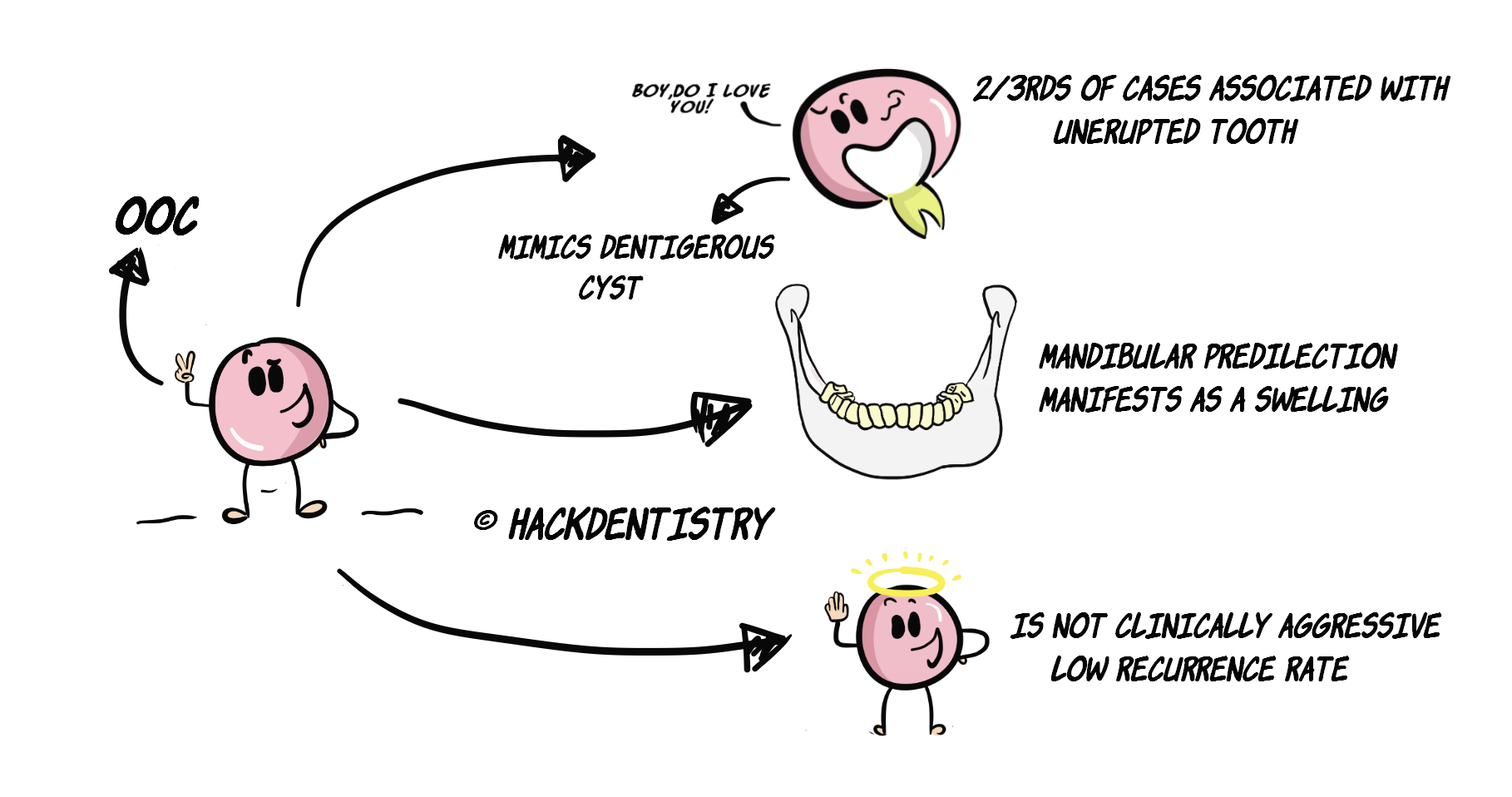
Almost 2/3rds (68%) of OOC cases are associated with an unerupted tooth mimicking a dentigerous cyst in the radiograph.
It affects the mandible more than the maxilla (mand:max-->3:1) and occurs predominantly in the posterior areas.
OOCs can vary from being small to large lesions. However they are more often associated with swelling of the jaws as compared to OKCs.
OOC is not clinically aggressive and has a low rate of recurrence.
RADIOLOGY FEATURES
OOC does not have a characteristic radiographic appearance. It is predominantly (93%) a unilocular lesion.
Almost 2/3rds of OOC cases show a pericoronal radiolucency (mimics dentigerous cyst).
Remember, pericoronal radiolucency usually refers to a unilocular radiolucency surrounding the crown of the tooth. A pericoronal radiolucency, however can be multilocular (eg – ameloblastoma, unicystic ameloblastoma, CEOT).HISTOPATHOLOGY FEATURES
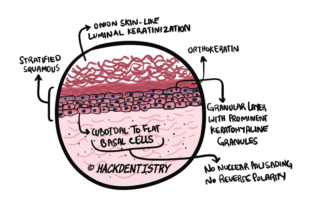
The epithelial lining in OOC is orthokeratinized stratified squamous and has onion-skin like luminal keratinization.
The epithelial lining has a granular layer with prominent keratohyaline granules and cuboidal to flat basal cells.
The epithelium- connective tissue interface is flat and has no rete ridges.
TREATMENT
OOC is not an aggressive lesion and is usually treated conservatively by enucleation and curettage.
QUESTIONS? BRING 'EM ON!!
Question 1:
Orthokeratinized odontogenic cyst is a variant of OKC. Is this true?
Answer:
Nope, OOC is not a variant of OKC!
Orthokeratinized odontogenic cyst (OOC) was first described as an orthokeratinizing variant of odontogenic keratocyst (OKC) in 1981. The basis of this distinction was the difference in its histopathology and its reduced chances of recurrence as compared to OKC.
It is now known and accepted that OOC is a distinct clinic-pathologic entityon its own. In factit has been listed as a developmental odontogenic cystin the recent (2017) WHO classification of odontogenic cysts.
Question 2:
Is OOC associated with Gorlin syndrome?
Answer:
Nope, it is not!
One of the distinctions between OOC and OKC is the latter’s association with Gorlin syndrome (Nevoid basal cell carcinoma syndrome). At least 90% of patients with Gorlin syndrome manifest with multiple OKCs. However there have not been records of any association of OOC with Gorlin syndrome.
✅HIGHLIGHTS - VIVA & ENTRANCE EXAM PERSPECTIVE
OOC is accepted to be clinico-pathologically different from odontogenic keratocyst(OKC) and is classified as an odontogenic cyst. It is not a variant of OKC.
Almost 2/3rds (68%) of OOC cases are associated with an unerupted tooth mimicking a dentigerous cyst in the radiograph.
OOC is not clinically aggressive and has a low rate of recurrence.
The epithelial lining in OOC is orthokeratinized stratified squamous and has onion-skin like luminal keratinization.
The epithelial lining has a granular layer with prominent keratohyaline granules.
📖REFERENCES AND FURTHER READING
MacDonald-Jankowski DS. Orthokeratinized odontogenic cyst: a systematic review. Dentomaxillofacial Radiology. 2010;39(8):455-467.
Speight PM, Takata T. New tumour entities in the 4th edition of the World Health Organization Classification of Head and Neck tumours: odontogenic and maxillofacial bone tumours. Virchows Arch. 2018 Mar;472(3):331-339.
Dong Q, Pan S, Sun LS, Li TJ. Orthokeratinized odontogenic cyst: a clinicopathologic study of 61 cases. Arch Pathol Lab Med. 2010 Feb;134(2):271-5.
Shear M, Speight PM. Cysts of the Oral and Maxillofacial Regions. 4 th ed. Blackwell Munksgaard; 2007.
Neville BW, Damm DD, Allen CM, Chi A. Oral and Maxillofacial Pathology. South Asian ed. Elsevier; 2016.
Regezzi JA, Sciubba JJ, Jordan RCK. Oral Pathology: Clinical Pathologic Correlations. 5 th ed. Elsevier; 2007.