
Salivary gland: Secretory cells and ducts (Note & Video

This video covers the classification, structure and the histology of the secretory cells and ducts of the salivary gland. I have split the whole video topic into two notes - one covering it's classification and structure and the other explaining the histology of the secretory cells and ducts.Salivary glands (SG) are compound, exocrine glands whose function is to secrete saliva.
Saliva, 90% of which is secreted by major salivary glands (which we shall discuss subsequently), is a multifunctional fluid, helping in maintaining the integrity of the oral cavity in various ways.
SECRETORY/ACINAR CELLS
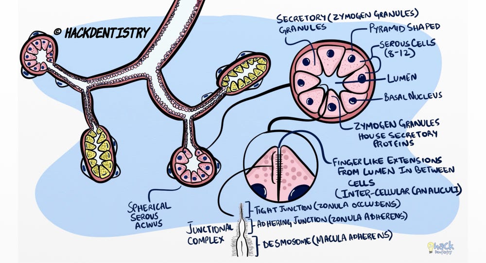
Serous cell
Serous cells are housed in a spherical acinus and may be 8-12 in number surrounding a central lumen.
A serous cell is pyramid shaped with a basal nucleus and numerous secretory granules in the apical cytoplasm.
These granules are called zymogen granules and house salivary macromolecules or proteins.
The lumen of the serous acinus has finger-like extensions that continue between the serous cells. These extensions are called canaliculi.
The cells are joined to each other on their lateral surfaces through junctional complexes. Each junctional complex is made of a tight junction (zona occludens), an adhering junction (zonula adherens) and a desmosome (macula adherens).
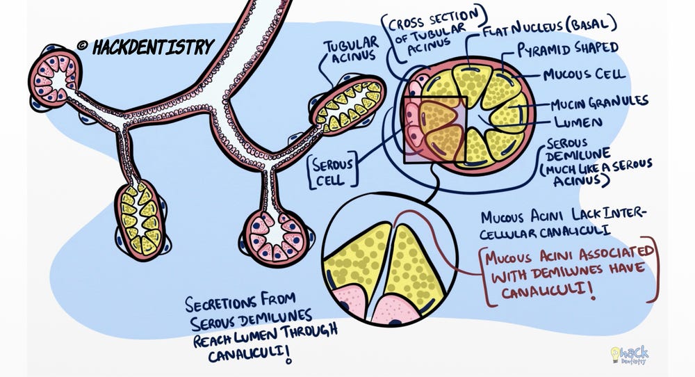
Mucous cell
Mucous acinar cells are also pyramidal cells with the apical cytoplasm packed with mucin granules and a flat nucleus in the basal cytoplasm compressed by mucin granules.
These cells are housed in tubular secretory end pieces and a cross section would show mucous acinar cells surrounding a central lumen.
Sometimes, mucous acini or end pieces may be associated with serous cells arranged on the them in the shape of a crescent. This is called a serous demilune.
Serous demilunes are much like serous acini.
Unlike, serous acini, mucous acini lack intercellular canaliculi.
However, those mucous acini associated with serous demilunes have canaliculi and the secretions of these serous demilunes reach the lumen of the end pieces through these canaliculi.
💡KNOW THY FACTS

Under the light microscope, the mucin granules/ mucin cannot be seen in H & E preparations, since they get washed out. Hence the cells look empty when stained with H & E stains. However, special stains like periodic acid-Schiff and alcian blue can stain mucin/mucus.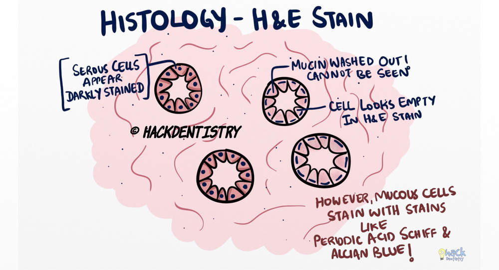
DUCTS AND DUCTAL CELLS
Intercalated duct
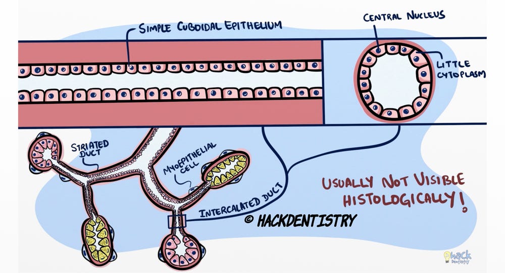
Secretory end pieces are continuous with the intercalated ducts.
Intercalated ducts are lined by simple cuboidal epithelium and are associated with myoepithelial cells.
The intercalated duct cells have a central nucleus and little cytoplasm.
Intercalated ducts are very small and are usually not visible under the microscope.
Several intercalated ducts join and open into striated ducts.
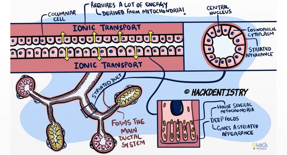
Striated duct
Striated ducts are lined by columnar cells which have a centrally placed nucleus and an eosinophilic cytoplasm.
The basal end of the striated ductal cell is thrown into numerous deep folds housing several mitochondria. These deep folds give the basal surface of the cell a striated appearance under the microscope, hence the name.
These ducts form the main portion of the salivary gland ductal system and are responsible for most of the ionic transport happening in the saliva when it travels from the end piece to the oral cavity.
This process requires a lot of energy and this is why these cells possess several mitochondria in the basal folds of their cytoplasm.
Excretory duct
Excretory ducts are present in the connective tissue septa between lobules and are called inter-lobular ducts.
Interlobular excretory ducts fuse to form bigger inter-lobar excretory ducts, with the main excretory duct opening into the oral cavity.
The inter-lobular excretory ducts are lined by pseudo-stratified columnar epithelium, and as the ducts get larger and approach the oral cavity, the lining becomes stratified squamous.
REFERENCES AND FURTHER READING
Berkovitz BKB, Hollan GR, Moxham BJ. Oral Anatomy, Histology and Embryology. 4th ed. Mosby Elsevier; 2009.
Nanci A. Tencate’s Oral Histology. Development, Structure and Function. 8th ed. Elsevier; 2013.
Kumar GS. Orban’s Oral Histology and Embryology.13th ed. Elsevier; 2011.
Avery JK. Oral development and Histology. 3rd ed. Thieme Medical Publishers; 2002.