
Salivary glands - Structure (Note & Video)

This video covers the classification, structure and the histology of the secretory cells and ducts of the salivary gland. I have split the whole video topic into two notes - one covering it's classification and structure and the other explaining the histology of the secretory cells and ducts.Salivary glands (SG) are compound, exocrine glands whose function is to secrete saliva.
Saliva, 90% of which is secreted by major salivary glands (which we shall discuss subsequently), is a multifunctional fluid, helping in maintaining the integrity of the oral cavity in various ways.
💡FUNCTIONS OF SALIVA

- The mucin in the saliva plays a major role in lubrication. This helps in prevention of oral tissues adhering to one another (keeps tissue moist). As a whole this helps in mastication, deglutition(swallowing) and speech.
- The buffering action of saliva, as a result of the presence of bicarbonate and phosphate ions, help in maintaining integrity of the teeth (protects from demineralization, promotes remineralization if demineralized).
- Proteins like lactoferrin and lysozyme in saliva have anti-bacterial action.
- Saliva helps in food digestion (amylase and lipase).
- Immunoglobulin A (IgA) is actively secreted in saliva. It contributes to mucosal immunity.
CLASSIFICATION OF SALIVARY GLANDS
SGs can be classified according to their size and the secretory cells they posses.
According to size
Major salivary glands
Minor salivary glands
Major salivary glands comprise 3 pairs of glands them being,
Parotid glands
Submandibular glands
Sublingual glands
Minor salivary glands are scattered throughout the oral cavity and are named according to their location.
Labial glands
Buccal glands
Palatal glands
Lingual glands (von Ebner’s glands)
Glossopalatine glands
Retromolar glands
💡KNOW THY FACTS

- The minor salivary glands are at least 600-1000 in number.
- The minor glands only contribute 10% of the total salivary secretion.
- Most minor salivary glands are predominantly mucous glands, with the exception of von Ebner's glands that are purely serous.
- Minor glands are not found on the gingiva and anterior hard palate.
According to type of “secretory cell”
SGs can be
Purely serous
Purely mucous
Mixed serous-mucous with either of the secretory cell predominating.
- Parotid glands are serous,
- Submandibular glands are mixed but are predominantly serous and
- Sublingual glands are also mixed but predominantly mucous.
- Majority of the minor salivary glands are mucous except for von Ebners glands which are serous glands.
STRUCTURE OF SALIVARY GLANDS
Secretory cells
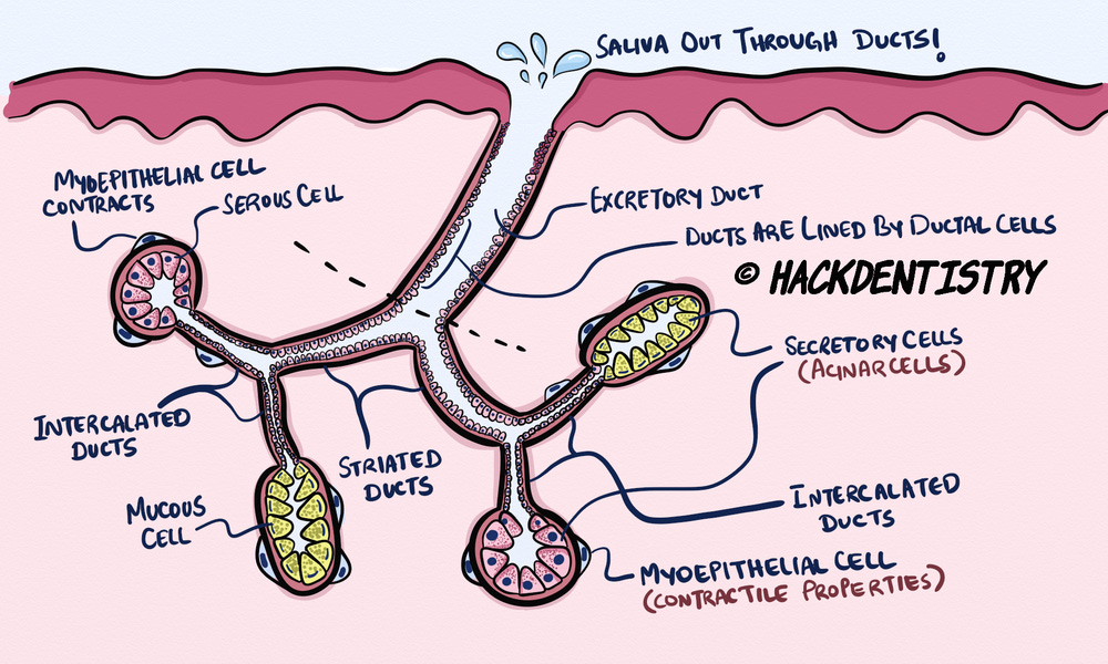
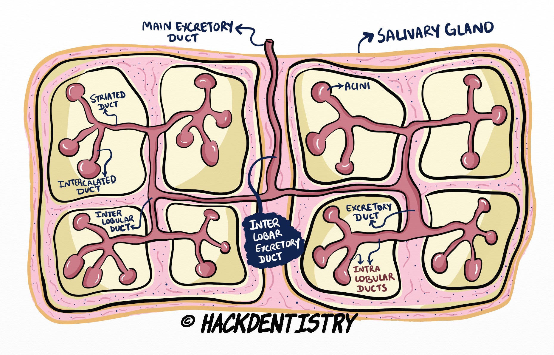
SGs are made of secretory units called “acini“ (singular - acinus), that house secretory cells.
Secretory cells are of two types - serous and mucous secretory cells.
Since secretory (serous and mucous) cells are housed in an acinus, the cells are also collectively called “acinar cells“.
The secretory units communicate with the oral cavity through a complex ductal arrangement.
The secretory cells (acinar cells) are responsible for secreting the primary salivary secretion.
Ductal arrangement
Secretory units open into an “intercalated duct“, which is continuous with the “striated duct“, which in turn opens into the “excretory duct“ of the salivary gland.
The ducts like the secretory units, are lined by cells called ductal cells.
The saliva secreted by acinar cells pass through the ducts to be excreted finally via the excretory duct into the oral cavity.
On it’s way, the saliva’s composition is influenced or affected by the intercalated and striated ducts.
Myoepithelial cells
The secretory end pieces and intercalated ducts are associated with stellate shaped contractile cells called the myoepithelial cells.
They are also called “basket cells” because they look like a basket holding a secretory unit.
Myoepithelial cells are epithelial cells that also have smooth muscle like contractile properties.
The main function of the myoepithelial cell is to contract and help squeeze out secretory products from the secretory end pieces to the ducts and finally to the oral cavity.
💡KNOW THY FACTS

This architecture of the salivary glands can be compared to a bunch of grapes, where the grapes represent the secretory end pieces (acini) and the stem/branches represent the ducts.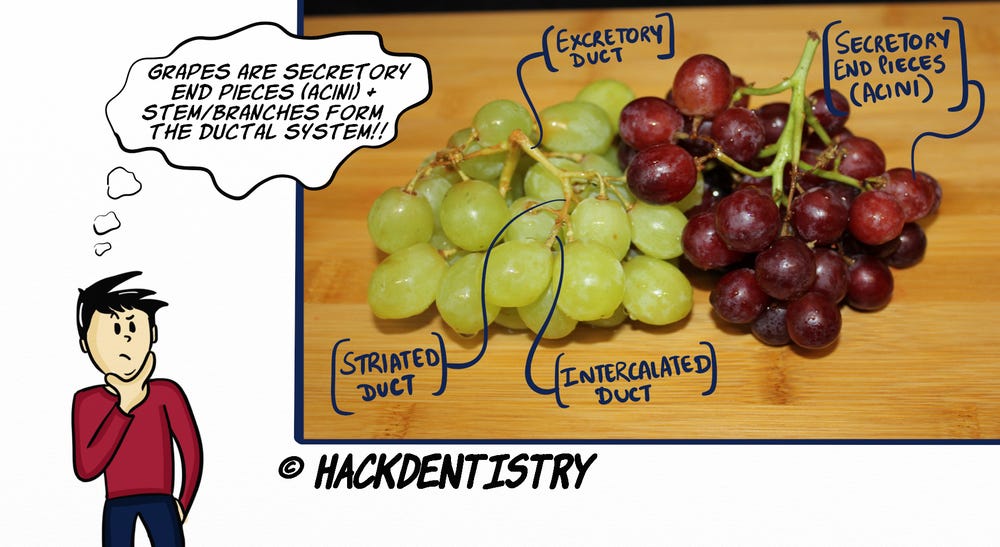
LOBULAR ARCHITECTURE
Salivary glands are basically enclosed by a connective tissue capsule that branches into inter-lobar septa (singular-septum) to divide the parenchyma/functional tissues into lobes.
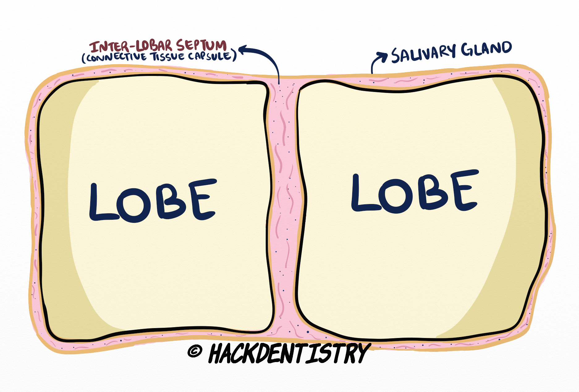
These lobes are further divided by inter-lobular septa into lobules.
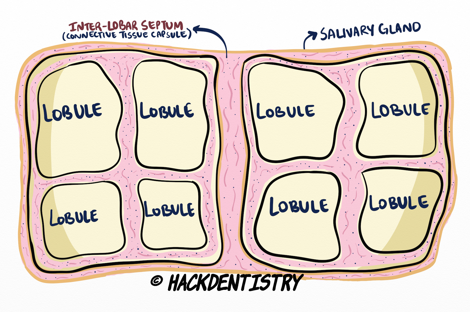
A lobule houses secretory end pieces, the intercalated and the striated ducts. Hence intercalated and striated ducts are called intra-lobular ducts.Striated ducts continue to open into excretory ducts in between lobules.
Excretory ducts in between lobules are inter-lobular excretory ducts. These continue and join other inter-lobular excretory ducts to form inter-lobar excretory ducts which in turn finally open into the main excretory duct of the salivary gland that is continuous with the oral cavity.
A NOTE ON ONCOCYTES
Oncocytes are cells that are packed with mitochondria and appear granular and deep pink under the microscope.
Oncocytes could appear in the acini and ducts of normal salivary glands.
The number of oncocytes in the salivary gland increase with age.
HIGHLIGHTS - VIVA & ENTRANCE EXAM PERSPECTIVE
SGs are made of secretory units called “acini“ (singular - acinus), that house secretory cells.
Secretory cells are of two types - serous and mucous secretory cells.
Since secretory (serous and mucous) cells are housed in an acinus, the cells are also collectively called “acinar cells“.
Secretory units open into an “intercalated duct“, which is continuous with the “striated duct“, which in turn opens into the “excretory duct“ of the salivary gland.
The ducts like the secretory units, are lined by cells called ductal cells.
The secretory end pieces and intercalated ducts are associated with stellate shaped contractile cells called the myoepithelial cells.
They are also called “basket cells” because they look like a basket holding a secretory unit.
Myoepithelial cells are epithelial cells that also have smooth muscle like contractile properties.
Major salivary glands comprise 3 pairs of glands them being, -> a) Parotid glands, b) Submandibular glands, c) Sublingual glands.
Parotid glands are serous, submandibular glands are mixed but are predominantly serous and sublingual glands are also mixed but predominantly mucous.
Majority of the minor salivary glands are mucous except for von Ebners glands which are serous glands.
REFERENCES AND FURTHER READING
Berkovitz BKB, Hollan GR, Moxham BJ. Oral Anatomy, Histology and Embryology. 4th ed. Mosby Elsevier; 2009.
Nanci A. Tencate’s Oral Histology. Development, Structure and Function. 8th ed. Elsevier; 2013.
Kumar GS. Orban’s Oral Histology and Embryology.13th ed. Elsevier; 2011.
Avery JK. Oral development and Histology. 3rd ed. Thieme Medical Publishers; 2002.