
Verrucous carcinoma (Note & Video)

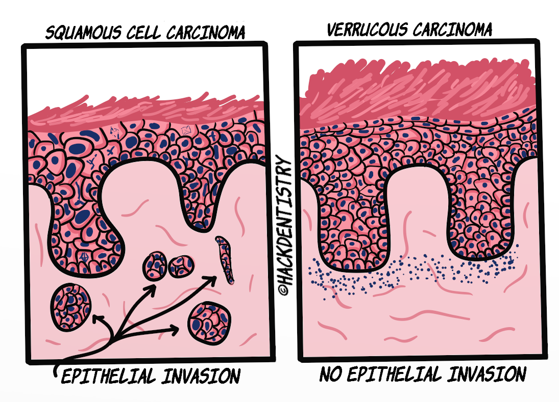
Verrucous carcinoma (VC) is a warty, exophytic variant of oral squamous cell carcinoma (OSCC).
Unlike OSCC, VC rarely exhibits regional or distant metastasis. VC however, is locally invasive and pushes into adjacent or surrounding tissues.
VC was described in detail by Ackerman in 1948 and is also called Ackerman’s tumor.
VC is usually associated with a smokeless tobacco/tobacco chewing habit. In fact, VC develops in the site where tobacco was placed and sometimes occurs in conjunction with submucous fibrosis in these patients.
It may also be associated with patients having smoking or drinking habits, poor oral hygiene, low socio-economic status or rarely no habits at all.
CLINICAL FEATURES
VC predominantly occurs in elderly males usually in the sixth decade.
It could potentially occur at any site in the oral cavity. Most common sites --> buccal mucosa, lower alveolar mucosa and vestibule, gingiva and tongue.
VC clinically appears large & diffuse, verrucous, warty or exophtic with distinct borders.
The lesion may be white or sometimes red-white in colour.
VC has a low propensity for metastasis but is locally invasive. If left untreated it could push and invade adjacent structures like periosteum, bone and salivary glands.
Enlarged lymph nodes in individuals with VC are due to secondary inflammatory changes and are not due to metastatis of the lesion.
VC could occur in conjunction with other oral potentially malignant disorders like leukoplakia and oral submucous fibrosis.
💡KNOW THY FACTS

Exophytic verrucous hyperplasia (EVH) is a verrucous lesion, known to occur in association with betel quid chewing that clinically and histopathologically resembles VC. However, EVH is distinguished histopathologically from VC by the absence of an endophytic growth pattern. Also, EVH though not always, may manifest with epithelial dysplasia.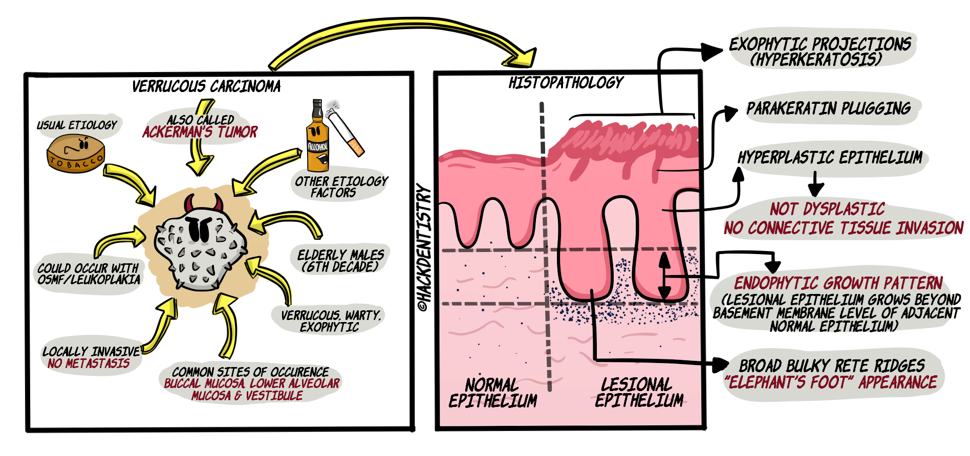
HISTOPATHOLOGY FEATURES
The epithelium has a verruciform or exophytic surface with clefts in between the exophtic projections.
The clefts are filled with parakeratin --> parakeratin plugging.
The epithelium is hyperplastic and is deceptively benign. It does not show dysplastic features (rarely manifests dysplasia).
Rete ridges are elongated, thick and bulky, and push into the adjacent connective tissue. The rete ridges are described as having an “elephant’s foot” appearance.
The characteristic feature of verrucous carcinoma is its endophytic growth pattern --> the lesional epithelium, with an intact basement membrane, pushes deep into the connective tissue (but does not invade the connective tissue like squamous cell carcinoma) and grows beyond the level of the basement membrane of the adjacent normal epithelium.
VC may show a dense inflammatory infiltrate in the connective tissue immediately below the epithelium.
💡CAUTION

- An adequate incisional biopsy is required for an accurate diagnosis of VC. Some amount of normal tissue is to be incised along with the lesional tissue. This is because normal epithelium is required for a comparison with the lesional tissue in order to establish the endophytic growth of VC (refer histopathology features).

- Also care must be taken to rule out any foci of squamous cell carcinoma (foci of lesional epithelium in the connective tissue) in the VC specimen.
TREATMENT
A complete surgical excision is the treatment of choice.
VC has an excellent prognosis with surgical management.
Patients are to be kept under follow-up as there are chances of recurrence of the lesion. Recurrence for VC has been reported in the range of 6-40% of the cases.
✅HIGHLIGHTS - VIVA & ENTRANCE EXAM PERSPECTIVE
Unlike OSCC, VC rarely exhibits regional or distant metastasis. VC however, is locally invasive and pushes into adjacent or surrounding tissues.
Enlarged lymph nodes in individuals with VC are due to secondary inflammatory changes and are not due to metastatis of the lesion.
VC could occur in conjunction with other oral potentially malignant disorders like leukoplakia and oral submucous fibrosis
The epithelium is hyperplastic and is deceptively benign. It does not show dysplastic features (rarely manifests dysplasia).
Rete ridges are elongated, thick and bulky, and push into the adjacent connective tissue. The rete ridges are described as having an “elephant’s foot” appearance.
The characteristic feature of verrucous carcinoma is its endophytic growth pattern.
REFERENCES AND FURTHER READING
Donald PM, Renjith G, Arora A. Tobacco Pouch Keratosis in a young individual: A brief description. J Indian Soc Periodontol. 2017;21(3):249–251.
Neville BW, Damm DD, Allen CM, Chi A. Oral and Maxillofacial Pathology. South Asian ed. Elsevier; 2016.
Rajendran R, Sivapathasundaram B. Shafer’s Textbook of Oral Pathology. 7th ed. Elsevier; 2012.
Regezzi JA, Sciubba JJ, Jordan RCK. Oral Pathology: Clinical Pathologic Correlations. 5 th ed. Elsevier; 2007.